
CURRY is an ideal software platform for combining and processing the various datasets that are obtained from a patient during an Epilepsy evaluation. CURRY combines multiple data sets (EEG, ECoG, MEG, MRI, fMRI, CT, DTI, and PET) to ensure the maximum information from the patients complete data set is utilized in making critical decisions.
CURRY uses a new interface, complete with automated workflow, data processing macros and import wizards to ensure the software is easy to work with. CURRY highlights for clinical applications are listed below.
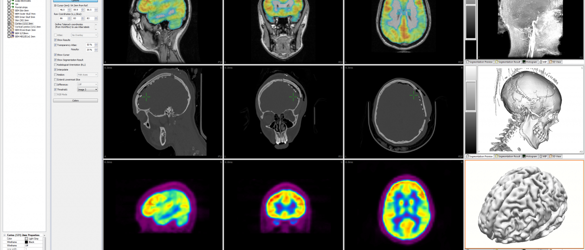
Image Co-Registration
One of the most powerful features of CURRY is its ability to merge multiple image datasets and allow critical features to be superimposed for optimal utility. For instance, electrode positions obtained from the CT can be superimposed on the MRI for validation of location accuracy. Similarly PET or fMRI activation can be superimposed on MRI to assist in determining areas of activation.
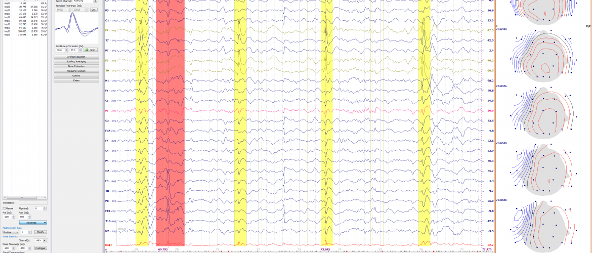
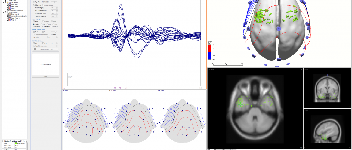
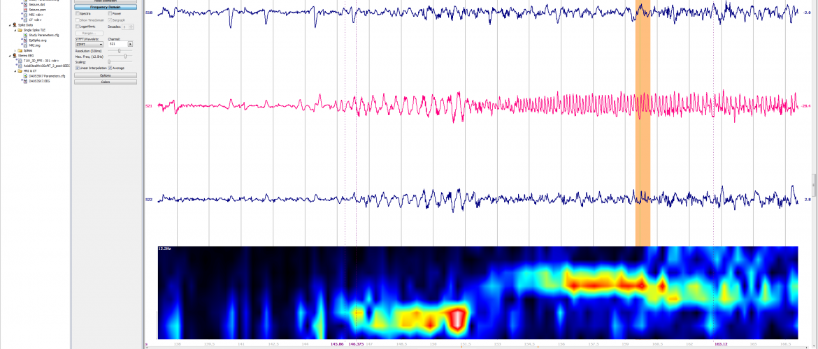
EEG Data Analysis and Mapping
CURRY has advanced analysis for surface recordings, including spike identification, averaging and statistical analysis. Subsequent source analysis can be applied to average spike activity or individual spikes (for clustering analysis). CURRY generates 2D and 3D topographic maps, based on individual anatomy obtained from the MRI.
Grid and Strip Planning and Visualization
Advanced modules within CURRY allow for grid and strip placement planning on a 3D rendering of the individual cortex including source results obtained from surface EEG. Grid and strip placement can later be identified and confirmed by a post-op CT.
File Format and Compatibility
CURRY seamlessly reads EEG data recorded from Compumedics and Neuroscan systems. CURRY imports EEG data from: EDF, ASCII, EGI, Micromed, Nicolet, Nihon Kohden, Persyst, Stellate, and XLTEK. CURRY reads MEG data from Elekta Neuromag, BTI, CTF, and Yokogawa. Contact us in case your EEG or MEG data format is not listed here.
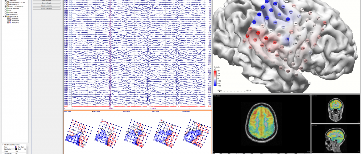
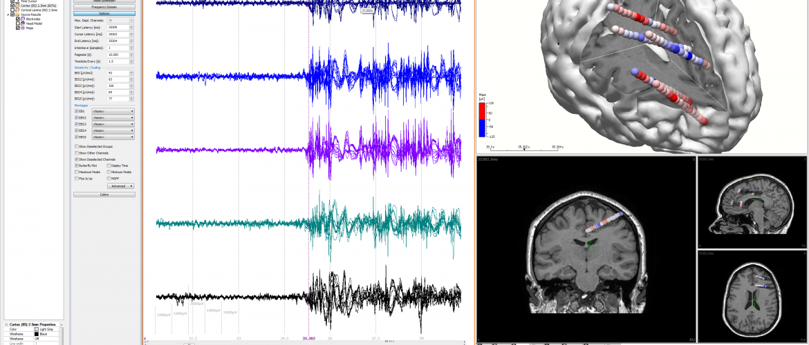
Grid, Strip and Stereo Electrode Locations
Based on the co-registered post-op CT images, the exact electrode coordinates are identified. Later, these will be used to map ECoG data, conduct source analysis and identify area of spike onset.
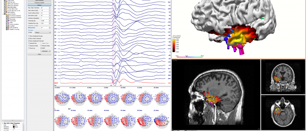
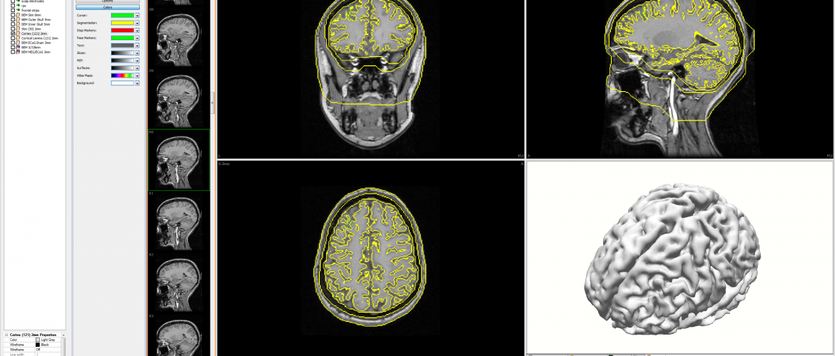
Functional Data in an Anatomical Context
CURRY provides for the functional data recorded as surface EEG or ECog to be mapped onto the cortical surface of the rendered cortex. This expression of the recorded functional data provides a clear picture of the activation patterns and area involvement derived from the recorded data.